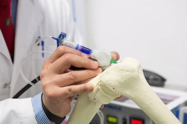
BioPen developed, uses 3D printing methods to 'draw' live cells onto patients
Press Trust of India, December 26, 2013

Doctors may soon be able to 'draw' new bone, skin and muscle on to patients, after scientists created a pen-like device that can apply human cells directly on to seriously injured people.
The device contains stem cells and growth factors and will give surgeons greater control over where the materials are deposited.
It will also reduce the time the patient is in surgery by delivering live cells and growth factors directly to the site of injury, accelerating the regeneration of functional bone and cartilage, scientists said.
The device developed at the University of Wollongong (UOW) will eliminate the need to harvest cartilage and grow it for weeks in a lab.
The BioPen works similar to 3D printing methods by delivering cell material inside a bio-polymer such as alginate, a seaweed extract, protected by a second, outer layer of gel material.
The two layers of gel are combined in the pen head as it is extruded onto the bone surface and the surgeon 'draws' with the ink to fill in the damaged bone section.
A low powered ultra-violet light source is fixed to the device that solidifies the inks during dispensing, providing protection for the embedded cells while they are built up layer-by-layer to construct a 3D scaffold in the wound site.
Once the cells are 'drawn' onto the surgery site they will multiply, become differentiated into nerve cells, muscle cells or bone cells and will eventually turn from individual cells into a thriving community of cells in the form of a functioning a tissue, such as nerves, or a muscle.
The device can also be seeded with growth factors or other drugs to assist regrowth and recovery, while the hand-held design allows for precision in theatre and ease of transportation.
The BioPen prototype was designed and built using the 3D printing equipment in the labs at Wollongong and was handed over to clinical partners at St Vincent's Hospital Melbourne, led by Professor Peter Choong, who will work on optimising the cell material for use in clinical trials.
"This type of treatment may be suitable for repairing acutely damaged bone and cartilage, for example from sporting or motor vehicle injuries," Choong, Director of Orthopaedics at St Vincent's Hospital Melbourne said.


No comments:
Post a Comment